Law enforcement technologyPortable, super-high-resolution 3-D imaging
A simple new imaging system could help manufacturers inspect their products, forensics experts identify weapons, and doctors identify cancers.
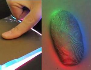
Super-high definition image by GelSight // Source: findbiometrics.com
By combining a clever physical interface with computer-vision algorithms, researchers in MIT’s Department of Brain and Cognitive Sciences have created a simple, portable imaging system that can achieve resolutions previously possible only with large and expensive lab equipment. The device could provide manufacturers with a way to inspect products too large to fit under a microscope and could also have applications in medicine, forensics, and biometrics.
The GelSight was demonstrated at the SIGGRAPH event last week in Vancouver, Canada.
In a 2009 paper, Edward Adelson, the John and Dorothy Wilson Professor of Vision Science and a member of the Computer Science and Artificial Intelligence Laboratory, and Micah Kimo Johnson, who was a postdoc in Adelson’s lab at the time, reported on an earlier version of GelSight, which was sensitive enough to detect the raised ink patterns on a $20 bill. At this year’s Siggraph — the premier conference on computer graphics — Adelson and Johnson, along with graduate student Alvin Raj and postdoc Forrester Cole, are presenting a new, higher-resolution version of GelSight that can register physical features less than a micrometer in depth and about two micrometers across.
Moreover, because GelSight uses multiple cameras to measure the rubber’s deformation, it can produce 3-D models of an object, which can be manipulated on a computer screen for examination from multiple angles.
Traditionally, generating micrometer-scale images has required a large, expensive piece of equipment such as a confocal microscope or a white-light interferometer, which might take minutes or even hours to produce a 3-D image.
The release notes that often, such a device has to be mounted on a vibration isolation table, which might consist of a granite slab held steady by shock absorbers. Adelson and Johnson, however, have built a prototype sensor, about the size of a soda can, which an operator can hold in one hand and which produces 3-D images almost instantly.
Adelson and Johnson are already in discussion with one major aerospace company and several manufacturers of industrial equipment, all of whom are interested in using