Infectious diseaseSecrets of plague unlocked with stunning new imaging techniques
Sandia Labs researchers have developed a super-resolution microscopy technique that is answering long-held questions about exactly how and why a cell’s defenses fail against some invaders, such as plague, while successfully fending off others like E.coli
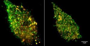
Sandia's new imaging technology (on right) with old // Source: sandia.gov
Researchers at Sandia National Laboratories have developed a super-resolution microscopy technique that is answering long-held questions about exactly how and why a cell’s defenses fail against some invaders, such as plague, while successfully fending off others like E.coli. The approach is revealing never-before-seen detail of the cell membrane, which could open doors to new diagnostic, prevention, and treatment techniques. “We’re trying to do molecular biology with a microscope, but in order to do that, we must be able to look at things on a molecular scale,” says Jesse Aaron, postdoctoral appointee at Sandia Labs.
The cell membrane is a bustling hub of activity on a miniscule scale. While providing structure and housing the cell’s interior, the membrane regulates movement of materials in and out of the cell, controls adhesion to other objects and coordinates the cell’s communications and subsequent actions through signaling. Receptor proteins on the surface of immune cells, known as toll-like receptors (TLRs), are tasked with recognizing intruders, or antigens. The TLR4 member of this receptor family responds to certain types of bacteria by detecting lipopolysaccharides (LPS) present on their surface. TLR4 proteins then alert the cell and activate an immune response.
Sandia Labs release reports that using imaging techniques they developed, Sandia researchers Aaron, Jeri Timlin, and Bryan Carson discovered that TLR4 proteins cluster in the membrane when confronted with LPS derived from E.coli, which increases cell signaling and response. Interestingly, LPS derived from the bacteria that cause plague, Yersinia pestis, do not cause the same effects. This could explain why some pathogens are able to thwart the human immune system.
The plague studies marked the first time such small events have been imaged and compared, the Sandia researchers said. Previously, even the most sophisticated optical microscopes could not image the cell surface with enough spatial resolution to see the earliest binding events, due to the diffraction barrier, which limits what can be resolved using visible light.